



CBCT vs Dental X Rays: Different Images Answer Different Questions
Dental imaging is not one size fits all. A small 2D X ray may be exactly what a dentist needs to check for a cavity between two teeth. A 3D cone beam computed tomography scan, commonly called CBCT, may be more helpful when the question involves bone, root anatomy, an impacted tooth, an implant site, or another structure that cannot be fully evaluated in two dimensions.
At Natural & Cosmetic Dentistry, the goal is not to choose the most advanced image for every patient. It is to choose the right image for the clinical question, using the smallest appropriate area and the lowest exposure that still provides diagnostically useful information.
This patient-specific approach is consistent with current dental imaging recommendations. The American Dental Association emphasizes that imaging should be based on clinical findings and individual need rather than performed routinely, and that CBCT should be incorporated when lower-exposure options cannot provide the necessary diagnostic information.
What Is a CBCT Scan?
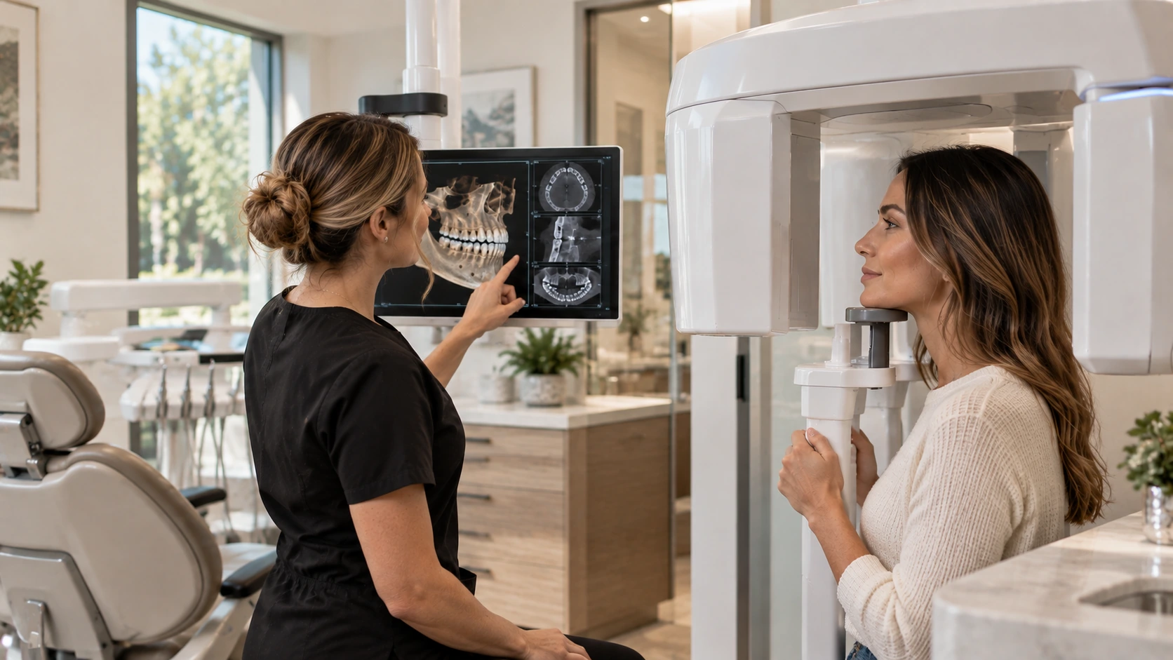
CBCT is a dental imaging system that uses a cone-shaped X ray beam while the scanner rotates around the head. Computer software then reconstructs the information into a three-dimensional view of the teeth, roots, jawbone, sinuses, airway space, and other nearby anatomy within the selected scan area.
Because the image can be viewed from multiple angles and in thin slices, CBCT can reveal spatial relationships that may overlap or remain hidden on a flat image. That additional information can help a dentist diagnose a complex problem and plan care with greater anatomical detail.
What Regular Dental X Rays Do Well
Traditional dental radiographs remain essential diagnostic tools. Bitewing, periapical, and panoramic images each answer different questions, and they generally involve less radiation than a CBCT scan.
- Bitewing X rays help detect decay between teeth and evaluate the height of the supporting bone.
- Periapical X rays show an entire tooth from crown to root and the bone around the root tip.
- Panoramic X rays provide a broad 2D overview of the jaws, teeth, and surrounding structures.
For routine exams and many focused concerns, a 2D image provides everything needed. CBCT does not replace these images. It adds a 3D option when a conventional X ray cannot answer the clinical question clearly enough.
CBCT vs Dental X Rays Side by Side
| Feature | Regular Dental X Rays | Dental CBCT |
|---|---|---|
| Image | 2D view | 3D reconstruction with multiple views and slices |
| Best suited for | Routine decay, bone levels, individual teeth, and broad screening | Questions involving 3D anatomy, location, depth, or proximity to nearby structures |
| Radiation | Usually lower than CBCT | Usually higher than 2D dental imaging, but generally lower than conventional medical CT |
| Selection | Often the first and most appropriate image | Used when the added 3D information is expected to change or clarify diagnosis or treatment planning |
The important point is that more imaging is not automatically better imaging. The most useful image is the one that answers the clinical question without exposing the patient to radiation that is unlikely to provide meaningful additional information.

When a Dentist May Recommend CBCT
A CBCT scan may be considered when the 3D view is likely to provide information that a clinical examination and lower-exposure images cannot provide. Depending on the patient and the question being evaluated, examples may include:
- Planning dental implant placement and evaluating available bone and nearby anatomy
- Locating impacted, unerupted, or unusually positioned teeth
- Evaluating complex root and canal anatomy or a persistent endodontic concern
- Assessing jawbone changes, facial trauma, or certain cysts and other findings
- Reviewing bony jaw joint anatomy when that information is clinically relevant
- Supporting selected orthodontic, airway, or dental arch evaluations when a 3D image is justified
The reason for the scan should always be specific. More detail is useful only when it helps answer a meaningful diagnostic or treatment-planning question.
How Green CBCT Technology Helps Limit Radiation
Our Green CBCT system is designed to produce diagnostically useful 3D images with dose-conscious protocols. One important feature is a selectable field of view. Instead of automatically scanning the largest possible area, the scan can be limited to the region that needs evaluation when the clinical situation allows. Optimized imaging protocols and short scan times can also help reduce unnecessary exposure and motion-related retakes.
An important distinction: Green does not mean radiation-free, and it does not mean every CBCT scan uses less radiation than a routine dental X ray. CBCT generally uses more radiation than conventional 2D dental radiographs, but less than conventional medical CT. Green CBCT technology helps reduce exposure within 3D dental imaging by matching the scan settings and field of view to the diagnostic need.
Is a CBCT Scan Safe?
CBCT uses ionizing radiation, so it should not be taken without a clinical reason. Current recommendations from the U.S. Food and Drug Administration and the American Dental Association emphasize selecting dental imaging according to the individual patient’s needs and minimizing unnecessary exposure. This includes using CBCT when the additional information is clinically justified rather than simply because 3D technology is available.
This dose-conscious approach is often described by the principle ALARA, meaning “as low as reasonably achievable.” Before recommending an image, the dentist considers factors such as the patient’s symptoms, medical and dental history, examination findings, previous images, age, disease risk, and the specific area requiring evaluation.
When CBCT is appropriately selected, the diagnostic benefit should outweigh the small risk associated with radiation exposure.
What to Expect During a Dental CBCT Scan
A dental CBCT is generally quick and does not require recovery afterward. The dental team will position you carefully before the scan so the area of interest can be captured accurately.
- You will be positioned carefully and asked to remain still.
- The scanner rotates around your head without touching you.
- The scan itself is brief, although positioning and setup take additional time.
- No special recovery is needed after the image is taken.
Tell the dental team if you are or may be pregnant, and let them know about any recent dental or medical imaging. Previous images may already contain some or all of the information needed, which can help avoid unnecessary additional imaging.
Frequently Asked Questions
Does CBCT replace regular dental X rays?
No. Regular dental X rays remain the preferred first choice for many common questions because they are efficient, highly useful, and usually involve less radiation. CBCT is reserved for situations where a 3D view adds information that matters to diagnosis or treatment planning.
Is CBCT lower radiation than an X ray?
It depends on what is being compared. A dental CBCT scan generally has a lower dose than conventional medical CT, but it usually has a higher dose than a routine 2D dental X ray. Actual exposure varies according to the machine, field of view, resolution, scan protocol, and patient.
Why not take a CBCT for every patient?
Because the added detail is not necessary for every examination and comes with additional radiation exposure. Current recommendations support patient-specific imaging rather than taking X rays or CBCT scans automatically on a fixed schedule.
Can CBCT find every dental problem?
No single image can answer every dental question. Diagnosis may require a clinical examination, dental history, photographs, pulp or bite testing, 2D X rays, CBCT, or a combination of these tools. The value comes from matching the diagnostic tool to the problem.
Why would my dentist need a 3D image of one tooth?
Sometimes a conventional X ray cannot clearly show the location or relationship of roots, bone, canals, or nearby anatomical structures. A limited-field CBCT may provide the additional perspective needed without scanning a larger area than necessary.
Should I bring my previous dental X rays to my appointment?
Yes, when they are available. Previous images may provide useful diagnostic information and can help the dentist determine whether new imaging is actually necessary.
The Right Image for the Right Reason
When comparing CBCT vs dental X rays, there is no single image that is automatically “better.” A traditional X ray may be all that is needed for one patient, while another dental concern may require the added anatomical detail of a 3D scan.
At Natural & Cosmetic Dentistry in Clearwater, Florida, we select imaging according to the clinical question. If a traditional X ray is sufficient, we use it. When 3D information is needed, our Green CBCT system can provide a more complete anatomical view while allowing the scan area and protocol to be tailored to what is necessary.
If you have been told that you may need a CBCT scan, our team can explain what the image is intended to show, why it is being recommended, and how the findings may affect your treatment options. Call (727) 888-6523 or contact our Clearwater dental office to schedule a comprehensive evaluation.
